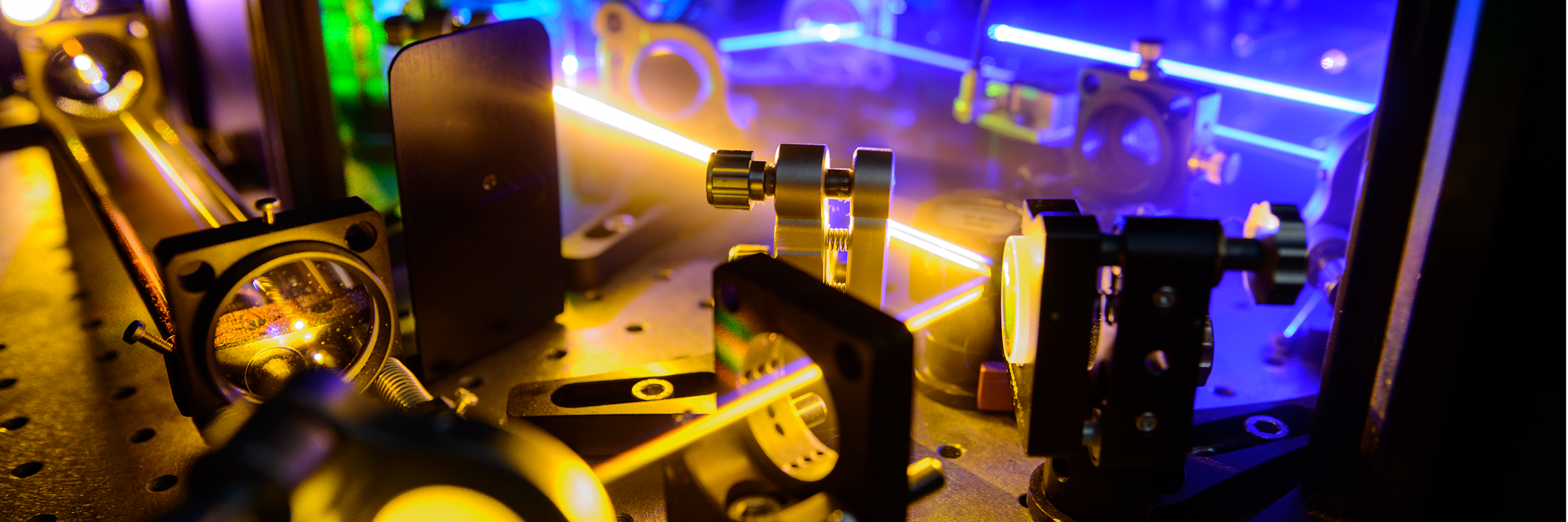
 Super-resolution microscopy allows to visualize structures in cells that were previously inaccessible. It is progressing at an amazing pace, thereby opening up entire new fields in biology. We use various super-resolution modalities including STED, RESOLFT and GSDIM microscopy, each of them exhibiting different strengths. For example, RESOLFT is especially suited for the imaging of living cells and STED for the fast recording of small areas. A major focus of our research is on the nanoscale architecture and dynamics of mitochondria. We also investigate new labelling strategies and are especially interested in the analysis and development of new fluorescent proteins for RESOLFT super-resolution microscopy.
References
Sahl, S.J., Hell, S.W. & Jakobs, S. : "Fluorescence nanoscopy in cell biology"
Nature Reviews Molecular Biology 18685-701, DOI 10.1038/nrm.2017.71Details
Danzl, J.G., Sidenstein, S.C., Gregor, C., Urban, N.T., Ilgen, P., Jakobs, S., Hell, S.W.: "Coordinate-targeted fluorescence nanoscopy with multiple off states"
Nature Photonics 10 (2), 122-128, DOI 10.1038/nphoton.2015.266Details
Jakobs, S., and Wurm, C.A.: "Super-resolution microscopy of mitochondria"
Curr. Opin. Chem. Biol. 20, 9-15, DOI 10.1016/j.cbpa.2014.03.019Details
Klar, T.A.,Jakobs, S.,Dyba, M.,Egner, A. and Hell, S.W.: "Fluorescence microscopy with diffraction resolution barrier broken by stimulated emission"
PNAS 97 (15), 8206-8210. DOI 10.1073/pnas.97.15.8206Details